


BIOMARKERS OF PREMATURE INFLAMMAGING: EPIGENETIC AGE, BIOLOGICAL FRAILTY AND MARKERS OF SENESCENCE (Narrative literature review)
Abaturov O.E., Samsonenko S.V., Makoviichuk O.A.
Summary. Abstract. Immunosenescence and associated inflammaging are key mechanisms driving the progression of juvenile idiopathic arthritis (JIA) and systemic lupus erythematosus (SLE) in children, leading to premature aging of the immune system, therapy resistance, and early development of comorbidities. Modern biomarkers, particularly epigenetic clocks, enable the detection of premature inflammaging and open prospects for personalized senotherapy in pediatric rheumatology. The aim of the study: to systematize modern data on key biomarkers of premature inflammaging in pediatric JIA and SLE, assess their diagnostic and prognostic value, and explore their potential for personalized therapy and comorbidity prevention. Materials and methods: narrative literature review with elements of systematic search conducted in accordance with PRISMA 2020 guidelines. The search was performed in PubMed/MEDLINE, Scopus, Web of Science, and Google Scholar for the period 2000–2026 (last search date: March 23, 2026). Key terms included: «premature inflammaging», «immunosenescence», «epigenetic clocks», «biological frailty», «cellular senescence», «SASP», «juvenile idiopathic arthritis (JIA)», «systemic lupus erythematosus (SLE)». A total of 1682 records were identified; after duplicate removal, 1039 remained; after title/abstract screening, 245 full-text articles were assessed; after full-text review, 92 articles were included (original studies, systematic reviews, meta-analyses). Excluded: conference abstracts, letters, editorials, and publications without full text. Data were extracted using a standardized form. Meta-analysis was not performed due to heterogeneity. Results. Premature inflammaging in JIA and SLE is characterized by accelerated epigenetic aging (GrimAge, GrimAge2, DunedinPACE), biological frailty (FI >0.20–0.25), accumulation of senescent cells (p16INK4a, p21CIP1, SA-β-gal), and SASP (IL-6, TNF-α, IL-8, MMP-3/9, CXCL8).First-generation epigenetic clocks (Horvath, Hannum) estimate chronological age, second-generation (PhenoAge, GrimAge2) assess mortality risk and comorbidities, and third-generation (DunedinPACE) measure the pace of aging. The most promising for children are PedBE (buccal swab) and GrimAge2 (incorporating hsCRP). Biological frailty is best evaluated using the deficit accumulation model (FI), which includes JADAS/SLEDAI, height Z-score, HOMA-IR, PWV/AIx, and PedsQL Fatigue; the Fried phenotype model is limited due to the lack of pediatric norms. Among the 12 hallmarks of aging (López-Otín, 2023), the most relevant are senescence (p16, SA-β-gal), SASP, impaired autophagy (↓LC3-II, ↑p62), dysbiosis, and mitochondrial dysfunction (cf-mtDNA). In JIA, local synovial senescence predominates, while in SLE, systemic senescence affects podocytes, endothelium, and microglia. Conclusions. Premature inflammaging is a key pathogenetic factor in the chronicity of JIA and SLE, therapy resistance, and early comorbidities. Comprehensive assessment of biomarkers (GrimAge2/DunedinPACE, FI, p16INK4a/SA-β-gal, SASP panel, cf-mtDNA) enables diagnosis of premature aging, risk stratification for complications, and prediction of treatment efficacy. Prospects include a transition to precision inflammaging medicine using senolytics, senomorphics, and modulators of autophagy/microbiota. Large pediatric cohorts are needed to validate threshold values and facilitate clinical implementation.
DOI: 10.32471/rheumatology.2707-6970.20999
UDC: 616.72-002-053.5-021.3+616-002.52]-06-07-091.8:576.3/.7:612.017(048.8)
Introduction
Immune system aging (immunosenescence) and associated chronic low-level inflammation (inflammaging) is one of the central mechanisms of progression of many chronic non-communicable diseases, including autoimmune diseases [31, 32]. In pediatric rheumatology, the phenomenon of prematural (accelerated) inflammaging attracts special attention in children and adolescents with juvenile idiopathic arthritis (JIA) and systemic lupus erythematosus (SLE), when the immune profile and biological age correspond to the characteristics of the immune status of the elderly [50, 69].
In these patients, inflammaging is not merely a background condition; rather, it acts as an independent pathogenic factor contributing to the persistence of chronic inflammation, resistance to conventional and biologic therapies, early development of comorbid conditions (including atherosclerosis, osteoporosis, growth retardation, nephrosclerosis, and cognitive impairment), and increased susceptibility to infections [87, 90].
Contemporary approaches to the assessment of premature inflammaging extend far beyond the measurement of classical acute-phase proteins (hs-CRP, fibrinogen, serum amyloid A). They involve a multi-level framework that combines:
1. assessment of biological age using epigenetic clocks (Horvath, GrimAge, DunedinPACE, GlycanAge) [33, 60];
2. quantitative assessment of biological frailty according to models of phenotype and accumulation of deficits [5];
3. Identifying signs of aging (12 Hallmarks) with a focus on senescence, autophagy disorders, dysbiosis, and chronic inflammation [58].
Such an integrated approach allows not only to state the presence of inflammaging, but also to assess its contribution to pathogenesis, predict resistance to therapy, and timely plan senotherapy strategies (senolytics, senomorphics, autophagy modulators, microbiota), which opens up the prospect of transition to precision medicine in pediatric rheumatology [33]. Despite the growth of data on inflammaging in adults, pediatric studies remain fragmentary. Premature inflammaging in children with JIA and SLE may explain early resistance to therapy and comorbidity, making it a promising target for senolytic/senomodifying strategies.
The aim of this article is to systematize current data on key biomarkers of premature inflammaging in JIA and SLE, analyze their diagnostic and prognostic value, and to evaluate their potential for application in personalized approaches to therapy and the prevention of comorbidities.
Materials and methods
The study was carried out in the format of a narrative literature review with elements of a systematic search, conducted in accordance with the recommendations of PRISMA 2020 (Preferred Reporting Items for Systematic Reviews and Meta-Analyses).
A comprehensive search of scientific publications was carried out in the PubMed/MEDLINE, Scopus, Web of Science Core Collection, and Google Scholar databases, covering the period from 2000 to 2026 (last search conducted on March 23, 2026). Combined queries with Boolean logical operators (AND, OR) and key terms were used: «prematurity inflammaging», «immunosenescence», «epigenetic clocks», «biological frailty», «cellular senescence», «SASP», «juvenile idiopathic arthritis (JIA)», «systemic lupus erythematosus (SLE)».
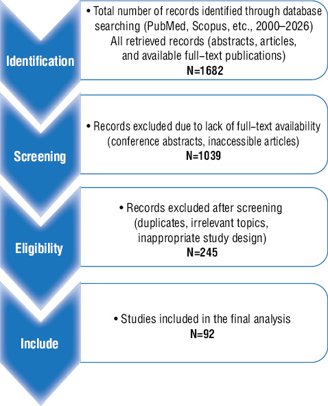
A total of 1682 records were identified. After removal of duplicates, 1,039 records remained. Following title and abstract screening, 245 full-text articles were assessed for eligibility. After full-text review, 92 studies (including original research articles, systematic reviews, and meta-analyses) were included in the final analysis. Conference abstracts, letters to the editor, editorials, and studies without available full text were excluded (figure).
The data were extracted in a unified form that included study design, sample characterization, disease type, biomarkers studied, molecular mechanisms, therapeutic approaches, and key findings. Due to the methodological heterogeneity of the studies, a meta-analysis was not performed. The generalization of the results was carried out by qualitative (narrative) synthesis.
The risk of systematic error of non-randomized trials (cohort, case-control, and mostly observational) was assessed using the Newcastle–Ottawa Scale (NOS), a standardized tool for assessing the quality of non-randomized trials. The scale evaluates three main domains:
- Selection (maximum 4 stars) — representativeness of the exhibited cohort, selection of the control group, establishment of exposure and lack of result at the beginning of observation;
- Comparability (maximum 2 stars) — control over the main confungi (age, gender, duration of the disease, concomitant therapy, etc.);
- Outcome/Exposure (maximum 3 stars) — reliability of endpoint determination, sufficiency of observation duration, and adequacy of follow-up.
The maximum rating is 9 stars. The results were interpreted as follows: 7–9 stars — low risk of bias, 5–6 stars — medium quality, ≤4 stars — low quality (high risk of bias). The assessment was carried out independently by the two authors, followed by a discussion of the discrepancies. NOS scores were considered in data interpretation and in formulating conclusions but were not used as exclusion criteria.
Research results
The most frequently cited characteristics of the included studies are provided in Table 1.
| № | Author, year | Country | Research design | Population (n, age) | Diseases | Key markers | Key results | Risk of bias | PMID |
|---|---|---|---|---|---|---|---|---|---|
| 1 | Horvath S., 2013 [40] | USA | Algorithmic modeling, validation | >8000 samples (various fabrics, age 0–100+ years) | General aging | 353 CpG sites (i.e. ELOVL2, FHL2, OTUD7A, ZYG11A, GPR158, PDE4C) | The first universal pan-fabric watch; correlation with chronological age r > 0.96; works in the blood, brain, skin, buccal epithelium | Low | 24138928 |
| 2 | Hannum G. et al., 2013 [37] | USA | Algorithmic Modeling + Validation | 656 people (blood, ages 19–101) | General aging | 71 CpG sites (mostly in the blood, associated with immune and metabolic genes) | Blood clock; high sensitivity to inflammatory markers (IL-6, CRP) | Low | 23177740 |
| 3 | Levine M.E. et al., 2018 [54] | USA | Algorithmic Modeling + Validation | >10,000 people (blood + 9 clinical biomarkers) | Phenotypic aging | 513 CpG + 9 biomarkers (albumin, creatinine, etc.) | PhenoAge is a better predictor of disease risk and mortality than Horvath | Low | 29676998 |
| 4 | Lu A.T. et al., 2022 [59] | USA | GrimAge improvements | Large cohorts (blood + biomarkers) | Prediction of mortality and the rate of aging | 1030 CpG + advanced biomarkers (HbA1c, CRP, telomeres) | GrimAge2 — even more accurately predicts mortality and the rate of aging | Low | 36516495 |
| 5 | Lu A.T. et al., 2019 [60] | USA / International | Algorithmic Modeling + Validation | >13,000 people (blood + 7 plasma markers) | Mortality prognosis and healthspan | 1030 CpG + 7 biomarkers (CRP, GDF-15, PAI-1, etc.) | GrimAge is the highest predictive accuracy for mortality, CVD, cancer | Low | 30669119 |
| 6 | Belsky D.W. et al., 2022 [4] | New Zealand / United States | Longitudinal cohort + algorithmic | Dunedin Study (n=1037, age 45 years at the time of analysis) | Aging rate | ~1000 CpG (dynamic, 173 key) | DunedinPACE — measures the rate of aging (years per year); >1.0 = acceleration | Low | 35029144 |
| 7 | McEwen L.M et al., 2020 [62] | Canada/International | Algorithmic Modeling + Validation | Children and adolescents (0–20 years, buccal swab) | Pediatric age | 94 CpG (buccal epithelium) | PedBE is the most accurate for non-invasive samples in children; easy to assemble | Low | 31611402 |
| 8 | Knight A.K., Smith A.K., 2016 [47] | USA | Algorithmic Modeling + Validation | Newborns (umbilical cord blood) | Gestational age | 148 CpG (cord blood) | Knight clock is the gold standard for estimating gestational age | Low | 27089367 |
| 9 | Bohlin J. et al., 2016 [7] | Norway | Algorithmic Modeling + Validation | Newborns (umbilical cord blood) | Gestational age | 96 CpG (cord blood) | Bohlin clock — very high accuracy for GA | Low | 27717397 |
| 10 | Lee Y. et al., 2019 [52] | Canada | Algorithmic Modeling + Validation | Placenta | Gestational age by placenta | ~100–200 CpG (placenta) | Lee clock is best for placental specimens | Low | 31235674 |
Notes: The Newcastle–Ottawa Scale does not apply to methodological/algorithmic work (all of which are not clinical case control or cohort studies).
The high level of evidence and low risk of bias of most studies (mainly methodological and validation works) confirm the reliability of epigenetic clocks as tools for early diagnosis, risk stratification and monitoring of the course of diseases. However, for widespread clinical adoption in pediatrics, large prospective cohorts of children with autoimmune diseases are required to refine threshold values and to validate pediatric-specific models.
Discussion
1. Epigenetic clocks: estimating biological age from DNA methylation
Epigenetic clocks (EpiC) allow you to determine the biological age of the immune system and the whole organism, which directly reflects the rate of senescence. Epigenetic clocks are based on the analysis of the level of DNA methylation in certain CpG sites of the genome, especially those that determine the development of an organism, for example, CpG sites near the Hox genes (homeobox) and Polycomb classes. studies have shown the relevance of EpiC to disease risk factors [21, 35, 47, 83]. DNA methylation is the covalent addition of a methyl group of DNA methyltransferases (DNMT) to the fifth carbon of cytosine, which is located next to guanine (CpG dinucleotides). DNA methylation, without altering the nucleotide sequence of DNA, modulates the availability of DNA to the transcriptional apparatus [42]. Methylated CpG dinucleotide is a stable epigenetic label associated with reduced gene expression. Changes in the DNA methylation landscape are associated with age [14] and the development of chronic diseases [48, 80, 88]. DNA methylation (DNAm) levels are evaluated using Illumina Infinium microarray technologies. For the analysis, Infinium HumanMethylation27 BeadChip (27 thousand CpG sites), Infinium HumanMethylation450 BeadChip (450 thousand sites) and the most modern Infinium MethylationEPIC (EPIC, covering more than 850 thousand sites) panels are used [36, 82].
Remodeling of the DNA methylation landscape forms the basis of epigenetic age [38, 92]. Children and adults whose epigenetic age i s older than chronological age are defined as individuals with positive epigenetic age acceleration (PEAA), and at epigenetic age younger than chronological age, negative epigenetic age acceleration (NEAA) is recognized [85]. Estimation of biological age by blood or tissue DNA methylation profiles allows you to assess both the rate of aging and the nature of the course of the disease or the effects of treatment. For example, the possibility of using EpiC in assessing the effectiveness of metformin treatment of type II diabetes mellitus has been shown [55].
1.1. Characteristics of the epigenetic clock
To date, several EpiCs have been created, which, according to Cynthia D J Kusters and Steve Horvath [47], can be represented by three generations (Table 2).
| Godinnik | Year of publication | Number of CpG sites | Data Type/Fabric | Main goal / advantage | Strengths | Weaknesses / limitations | Source |
|---|---|---|---|---|---|---|---|
| First generation | |||||||
| Horvath clock | 2013 | 353 | Pan-tissue, universal (blood, brain, skin, buccal epithelium, etc.) | Universal assessment of biological age in different tissues | Highest correlation with chronological age (r > 0.96), works in many tissues | Less sensitive to inflammation and immunosenescence than blood models | [40] |
| Hannum clock | 2013 | 71 | Whole Blood | Assessment of blood age, sensitivity to immune changes | Simple, high sensitivity to inflammatory markers (IL-6, CRP) | Limited by blood, works worse in other tissues | [37] |
| Second generation | |||||||
| PhenoAge | 2018 | 513 | Blood + 9 clinical biomarkers | Prognosis of phenotypic aging and mortality | Better predicts the risk of disease and mortality than Horvath | Designed for adults; poor accuracy in children; requires additional blood tests | [54] |
| GrimAge | 2019 | 1030 (Composite) | Blood +7 plasma markers | Best Mortality and Health Prognosis (healthspan) | Highest predictive accuracy (mortality, CVD, cancer); takes into account inflammation | Complex (many markers), requires expensive panels | [60] |
| GrimAge2 | 2022 | 1030 | Blood + biomarkers | Improved version of GrimAge with telomere length | Even more accurately predicts mortality and the rate of aging | Very complex; requires the maximum amount of data; poorly validated in children | [59] |
| Third generation | |||||||
| DunedinPACE | 2020 / 2022 | ~1000 (dynamic) | Blood (longitudinal data) | Pace of aging, years per calendar year | Measures the rate of aging (1.0 = normal, >1.0 = acceleration) | Requires longitudinal data for calibration, less common | [4] |
| Epigenetic clocks for use in children | |||||||
| PedBE (Pediatric Buccal Epigenetic) | 2020 | 94 | Buccal epithelium (buccal smear) | Best for non-invasive specimens in children (0–20 years) | Highest accuracy for buccal samples in children; easy to assemble | Limited only to buccal cells; not validated for blood/brain | [63] |
| Knight clock | 2016 | 148 | Umbilical cord blood | Gestational age in newborns | The gold standard for estimating gestational age in newborns | Limited only to umbilical cord blood; not for older children | [46] |
| Bohlin clock | 2016 | 96 | Umbilical cord blood | Gestational age in newborns | Very high accuracy for GA | Umbilical cord blood only; not for children after birth | [7] |
| Lee clock | 2019 | not a decree. (about 100–200) | Placenta | Gestational age by placenta | Best for Placental Specimens | Limited by the placenta; not for other fabrics | [52] |
| PhenoAge | 2018 | 513 | Blood | Phenotypic age (health/mortality) | Better predicts disease and mortality than Horvath clock | Poor accuracy in children; designed for adults | [13] |
Note: most models are developed in adult or mixed cohorts; pediatric validations are limited (especially <2 years); small study samples; sensitivity to inflammation and therapy (glucocorticoids, cytostatics) may skew the results.
It has been demonstrated that first-generation EpiCs (e.g., Horvath’s clock, Hannum’s clock) diagnose biological age and predict chronological age. However, they are less sensitive to predicting changes in human health.
At the same time, second-generation EpiCs (e.g., PhenoAge, GrimAge) predict healthy life expectancy and the risk of premature death. They better reflect the «wear and tear» of the body and directly correlate with the levels of pro-inflammatory factors (CRP, IL-6, TNF-α). It has been shown that inflammation is accompanied by hypomethylation of pro-inflammatory genes, for example, promoters of interleukin 6 (IL-6) genes, tumor necrosis factor alpha (TNF-α), which makes them constantly active [49, 75, 77].
The third generation of EpiC (e.g. DunedinPACE) estimates the rate of aging («pace of aging») in real time [47].
The most promising for pediatric rheumatology are PedBE (non-invasive, buccal smear, high accuracy in children 0–20 years old) and GrimAge2 (takes into account hsCRP and telomeres, ideal for evaluating inflammaging). DunedinPACE allows you to estimate the rate of aging over the past 12 months, which is especially valuable for monitoring JIA and SLE activity. For clinical implementation, large prospective pediatric cohorts are required to refine threshold values and validate age-appropriate models.
1.2. Recommendations for the use of EpiC in the diagnosis of inflammaging
To assess biological age in children, PedBE (buccal smear cells), Knight, Bohlin (tumbler blood cells), Lee (placental cells) are considered the best EpiC [86]. For the prognosis of disease or mortality in children, it is recommended to use GrimAge or PhenoAge with caution, as they are predominantly validated in adults [26]. Small study samples, limited number of tissues studied, poor EpiC validity in children younger than 2 years of age or in sick people (e.g., patients with JIA, SLE) significantly limit the use of epigenetic clocks. Autoimmune diseases JIA, SLE are accompanied by significant changes in the landscape of DNA methylation, which can be used to determine biological age to confirm the presence of inflammaging [12, 23, 28, 39, 51, 72]. We believe that epigenetic age can be one of the markers of the development of inflammaging, as a process associated with senescence.
For the introduction of epigenetic testing into clinical practice (in particular in JIA and SLE), it is recommended to use second-generation EpiC (GrimAge) to assess the risk of organ damage. The DunedinPACE and GrimAge epigenetic clocks are likely to become a tool for diagnosing inflammaging. However, the use of GrimAge and DunedinPACE in pediatric rheumatology is still limited due to the lack of validated studies of pediatric cohorts. The epigenetic clock of GrimAge2 [59] includes, as a component of the algorithm, levels of highly sensitive C-reactive protein (hsCRP), which makes it the most relevant for the diagnosis of inflammaging. At the same time, DunedinPACE measures «biological aging over the past 12 months», which is ideal for assessing JIA and SLE activity [4].
Thus, EpiC GrimAge, DunedinPACE are the most promising tools for assessing inflammaging in pediatric rheumatology, but their clinical application requires large prospective studies in children with JIA and SLE. To increase the accuracy of inflammaging diagnosis, it is advisable to use a combination of DunedinPACE (aging speed rate) and GrimAge2 (likelihood of complications risk), which will personalize the intensity of targeted therapy. It is recommended: 1) to start with PedBE (buccal smear) to assess biological age in children; 2) use GrimAge2 with DunedinPACE to assess inflammaging and the risk of comorbidities; 3) repeat measurements every 6 to 12 months in patients with active JIA/SLE to monitor the rate of aging and the effectiveness of therapy.
2. Systemic clinical and metabolic markers of «biological frailty» in children
The concept of biological frailty in the pediatric population remains an insufficiently standardized term. Frailty is defined as a state of increased vulnerability due to a decrease in physiological reserve and impaired ability to withstand stressors, leading to rapid deterioration in health, more frequent complications, and mortality [11, 24]. Biological frailty in children is manifested not by classical geriatric signs, but by a complex of delayed physical and sexual development, the appearance of metabolic disorders, a decrease in physical endurance and early manifestation of comorbid conditions [5, 11]. The conceptualization of biological frailty is especially important for children with chronic inflammatory and autoimmune diseases (JIA, SLE), disabilities, or complex care needs [10].
The Fried Frailty Phenotype model (Fried Frailty Phenotype, 2001) is the most common model in geriatrics and rheumatology [34]. Frailty is defined by the presence of ≥3 of the following 5 clinical signs:
1. unintentional weight loss (>4.5 kg or ≥5% of body weight per year),
2. feeling exhausted and tired (self-esteem on the CES-D scale or similar),
3. weakness (reduced strength of the hand, measured with a dynamometer),
4. low physical activity (according to questionnaires or energy expenditure <383 kcal/week in men and <270 kcal/week in women),
5. slow gait speed (time to walk 4 meters >6 seconds or according to age-specific norms).
This model is well validated in elderly patients and is associated with inflammaging (elevated IL-6, TNF-α, hs-CRP), senescence, and decreased muscle mass (sarcopenia). In children and adolescents with JIA and SLE, the frailty phenotype model is not used due to the lack of standardized pediatric norms, but individual signs (exhaustion, low physical activity, growth retardation) are often recorded as manifestations of prematural inflammaging.
According to the Frailty Index / Deficit Accumulation model, frailty is defined as the proportion of accumulated deficits (diseases, symptoms, dysfunctions, laboratory abnormalities) from the total number of possible deficits (usually 30–70 indicators) [63]. The Frailty Index (FI) is calculated using the formula FI = the number of existing deficiencies / the total number of deficits assessed. Frailty is diagnosed at FI >0.25; and severe frailty is diagnosed at FI >0.35–0.40.
This model is more flexible and applicable in pediatrics, it allows the inclusion of deficits specific to JIA and SLE:
1. disease activity (according to the JADAS-71, SLEDAI scales),
2. organ damage (nephritis, uveitis, carditis),
3. delayed physical and sexual development,
4. anemia of a chronic disease,
5. osteoporosis,
6. cognitive impairment, depression, low tolerance to physical activity,
7. frequent infections due to immunosuppression.
The frailty index in adults with rheumatic diseases and SLE often exceeds 0.25–0.30, which indicates prematurity frailty, possibly associated with chronic inflammaging [29, 50, 76, 81, 89].
A comparison of models for assessing biological frailty is provided in Table 3.
| Characteristics | A model of the frailty phenotype [34] | Deficit Accumulation Model (Frailty Index) [63] | Pediatric models [5] |
|---|---|---|---|
| Basic principle | ≥3 of 5 phenotypic criteria | Proportion of accumulated deficits | Adaptation of deficiencies or phenotypes to children’s norms |
| Number of criteria / deficits | 5 fixed criteria | 30 to 70 plus variables | 20–50 variables (often include JADAS/SLEDAI, Z-score) |
| Main criteria | Weight loss, exhaustion, weakness, low activity, slow gait | Diseases, symptoms, laboratory abnormalities, functional disorders | Growth retardation, bone age, HOMA-IR, PedsQL Fatigue, JADAS/SLEDAI, PWV |
| Brittleness thresholds | ≥3 criteria = frailty; 1–2 = frailty | FI >0.25 = brittleness; >0.35–0.40 = severe brittleness | FI >0.20–0.25 (pediatric cutoff lower); There is no single standard |
| Validation in pediatrics | Very low (no norms for children) | Moderate–high (flexible adaptation) | Moderate (individual components are validated: Z-score, PedsQL) |
| Benefits in pediatrics | Simplicity, clinical accessibility | Flexibility, takes into account specific childhood deficits | Best reflects pediatric manifestations (growth retardation, disease activity) |
| Disadvantages in pediatrics | Many criteria are not adapted (gait, hand strength) | Requires ≥30 variables for reliability; Time-consuming | Lack of a single standard; requires validation in JIA/SLE |
| Connection with Inflammaging | Moderate (IL-6, CRP, sarcopenia) | High (cumulative effect of inflammation) | High activity levels (JADAS/SLEDAI and metabolic markers) |
| Recommended use in JIA/SLE | Limited (select components only) | Recommended as the main model | Most Promising (Adapted FI) |
| Characteristics | A model of the frailty phenotype [34] | Deficit Accumulation Model (Frailty Index) [63] | Pediatric models [5] |
| Basic principle | ≥3 of 5 phenotypic criteria | Proportion of accumulated deficits | Adaptation of deficiencies or phenotypes to children’s norms |
| Number of criteria / deficits | 5 fixed criteria | 30 to 70 plus variables | 20–50 variables (often include JADAS/SLEDAI, Z-score) |
| Main criteria | Weight loss, exhaustion, weakness, low activity, slow gait | Diseases, symptoms, laboratory abnormalities, functional disorders | Growth retardation, bone age, HOMA-IR, PedsQL Fatigue, JADAS/SLEDAI, PWV |
| Brittleness thresholds | ≥3 criteria = frailty; 1–2 = frailty | FI >0.25 = brittleness; >0.35–0.40 = severe brittleness | FI >0.20–0.25 (pediatric cutoff lower); There is no single standard |
| Validation in pediatrics | Very low (no norms for children) | Moderate–high (flexible adaptation) | Moderate (individual components are validated: Z-score, PedsQL) |
| Benefits in pediatrics | Simplicity, clinical accessibility | Flexibility, takes into account specific childhood deficits | Best reflects pediatric manifestations (growth retardation, disease activity) |
| Disadvantages in pediatrics | Many criteria are not adapted (gait, hand strength) | Requires ≥30 variables for reliability; Time-consuming | Lack of a single standard; requires validation in JIA/SLE |
| Connection with Inflammaging | Moderate (IL-6, CRP, sarcopenia) | High (cumulative effect of inflammation) | High activity levels (JADAS/SLEDAI and metabolic markers) |
| Recommended use in JIA/SLE | Limited (select components only) | Recommended as the main model | Most Promising (Adapted FI) |
Note: FI thresholds >0.25–0.30 are adapted from adult studies; in children, pediatric adaptations often use lower thresholds (FI >0.20–0.25) or specific sets of deficits. Further validation studies are needed to establish thresholds for pediatric cohorts.
In JIA and SLE in children, both models of frailty can be applied, but the model of accumulation of deficits is more practical and sensitive, since it allows you to take into account specific childhood manifestations (growth retardation, nephritis, anemia, cognitive impairment) and their connection with premature inflammaging. It is this model that better reflects the cumulative effect of chronic inflammation, senescence and therapeutic burden on the biological age of the child.
2.1. Assessment of delayed physical development and puberty
Delayed linear growth and impaired pubertal development are among the most sensitive clinical markers of premature senescence and, potentially, inflammaging in children with chronic inflammatory diseases [1, 69].
A decrease in body length Z-score of ≥–2 SD over ≥6–12 months, delayed sexual development, and acceleration of bone age correlate with high levels of pro-inflammatory cytokines in JIA patients [25, 68].
In girls with SLE, delayed puberty is more common with high levels of IFN-α and IL-6 [17].
Regular monitoring of Z-score of body length, bone age (every 6–12 months) can allow the detection of children at high risk of premature inflammaging even at the preclinical stage of comorbid conditions.
2.2. Metabolic age and metabolic profile disorders
Children with JIA are more likely to experience metabolic disorders such as dyslipidemia, increased insulin resistance, and abnormal distribution of body fat compared to healthy peers. These disorders resemble early signs of metabolic syndrome and are considered to contribute to premature cardiovascular morbidity [43, 66]. Metabolic changes in children are often early manifestations of premature systemic aging, telomere shortening [16, 44, 67], and possibly inflammaging.
Dyslipidemia. Almost half of children with SLE have dyslipidemia, especially with early onset of the disease and a higher body mass index [66]. An increase in triglycerides, cholesterol due to a high-fat diet (High Fat Diet) correlates with the expression of the key marker of senescence p16INK4a, which promotes the accumulation of lipids in macrophages and enhances the activity of inflammation (inflammaging) [32, 56].
Insulin resistance. JIA and SLE are characterized by increased levels of HOMA-IR and decreased insulin sensitivity. Glucose metabolism disorders are associated with high levels of IL-6, TNF-α and inflamation surrogates [19, 71, 79]. In systemic JIA, insulin resistance often precedes the clinical manifestations of metabolic syndrome [43].
2.3. Functional indices as clinical surrogates for inflamation
One of the most important manifestations of premature inflammaging in JIA and SLE is a decrease in physiological reserves, which is manifested by real functional disorders. That is why the assessment of muscle strength, endurance, vascular rigidity and quality of life becomes a valuable clinical surrogate for a chronic inflammatory process.
Muscle strength and endurance are particularly sensitive indicators. In patients with JIA and SLE, there is often a decrease in the results of the 30-second chair stand test, a simple and reproducible method that allows you to estimate the speed and number of repetitions [85]. Hand dynamometry (handgrip strength) demonstrates a significant decrease in strength in patients compared to healthy peers [8]. A decrease in these indicators by more than 1.5 standard deviations from the age norm is associated with the accumulation of senescent cells in muscle tissue, which is confirmed in adult populations and probably has a similar mechanism in children with chronic inflammation [30].
Another important marker of inflamation is vascular stiffness. It is associated with endothelial dysfunction and the development of atherosclerosis. The pulse wave propagation rate (PWV, m/s) and the augmentation index (AIx,%) increase already at the preclinical stage in JIA and SLE, reflecting the chronic effect of pro-inflammatory cytokines on the vascular wall [16; 78].
Integral scales objectively complement the data for assessing the functional state. The PedsQL Multidimensional Fatigue Scale allows you to quantify the severity of fatigue [6]. The general scale of the Pediatric Quality of Life Inventory (PedsQL) reflects a decrease in quality of life, which is closely related to chronic inflammation and senescence [PedsQL, official documentation, 41]. At the same time, the scales of disease activity — Juvenile Arthritis Disease Activity Score (JADAS) in JIA and SLE Disease Activity Index 2000 (SLEDAI) in SLE — are indirect clinical indicators of inflammaging, since persistent disease activity, even at a low level, is associated with the accumulation of senescent load [41, 65].
Thus, the combination of functional tests (dynamometry, stool rise test), vascular stiffness assessment (PWV, AIx), integral quality of life and fatigue scales (PedsQL, Fatigue Scale) makes it possible to detect premature inflammaging at the clinical level, when laboratory markers can still remain within normal limits. These indicators of biological frailty directly reflect the impact of chronic inflammation on the child’s daily activity and long-term prognosis.
2.4. Recommendations for the use of markers of «biological frailty» in the diagnosis of inflammaging in JIA and SLE in children
Biological frailty is one of the key clinical manifestations of premature inflammaging in children with JIA and SLE. Its assessment makes it possible to predict resistance to therapy, the risk of complications. The most significant markers of biological frailty in children with JIA and SLE today are:
1. frailty index, Z-score of growth, bone age;
2. disease activity indices;
3. markers of insulin resistance;
4. augmentation index and PWV.
With an FI value of >0.25, there is a suspicion of the presence of biological frailty and premature inflammaging.
It is recommended to annually assess: 1) Z-score of growth and bone age; 2) HOMA-IR; 3) dynamometry of the hand and the test «getting out of the chair»; 4) PWV/AIx (in adolescents); 5) PedsQL Fatigue along with JADAS/SLEDAI assessment. With an FI > 0.25, it is advisable to strengthen monitoring of the development of comorbid conditions. At the same time, it should be noted that the cutoff value of 0.20–0.25 remains debatable and requires further validation in pediatric cohorts.
3. Biomarkers of aging and cellular senescence in inflammaging
In 2023, Carlos López-Otín and sang. [58] provided an updated list of 12 signs of aging (Hallmarks of Aging) (Table 4).
| № | Sign of aging | Brief description | Main biomarkers | Definition Level | Clinical significance |
|---|---|---|---|---|---|
| 1 | Genomic instability | Accumulation of DNA damage (mutations, breaks, adducts) due to impaired DNA repair | γ-H2AX foci, 53BP1 foci, 8-OHdG (oxidized DNA), micronuclei, mutation rate in somatic cells | PBMC, tissues, flow cytometry | Increased in SLE due to interferon stress |
| 2 | Telomere depletion | Progressive shortening of telomere repeats of TTAGGG at each division | Telomere length (qPCR, Flow-FISH, Southern blot), T/S ratio, telomerase activity (hTERT expression) | PBMC, Leukocyte | Accelerated shortening of telomeres in JIA and SLE |
| 3 | Epigenetic changes | Changes in DNA methylation, histone modifications, miR expression | Epigenetic clocks (Horvath, GrimAge, DunedinPACE), global methylation level (5-mC), H3K9me3, H3K27me3 | Blood, PBMC (Illumina EPIC 850K) | Acceleration of epigenetic age in chronic diseases |
| 4 | Loss of proteostasis | Disorders of protein synthesis, folding, transport and degradation | Accumulation of aggregated proteins, ubiquitin levels, ↑ p62/SQSTM1, lipofuscin, amyloid aggregates, ↓ proteasomal activity | PBMC, tissues, Western blot | Increased in garb aging and SASP |
| 5 | Impaired nutrient sensitivity | Dysregulation of mTORS, AMPK, insulin/IGF-1, sirtuin (SIRT1) | mTORC1 activity (phosphorylation of S6K1, 4E-BP1), ↓ AMPK, ↓ SIRT1, IGF-1/ insulin signaling, NAD+ level | PBMC, serum | mTOR hyperactivation in JIA and SLE |
| 6 | Mitochondrial dysfunction | Decreased OXPHOS, ↑ ROS, mtDNA damage, mitophagy disorders | cf-mtDNA in plasma, ROSA (DCFH-DA), cardiolipin, ↓ PGC-1α, ↓ LC3-II (mitophagy), cytochrome c release | Plasma, PBMC, fluorescence | mtDNA leakage promoting IFN-I production in SLE |
| 7 | Cellular senescence | Persistent arrest of the cell cycle, formation of SASP | ↑ p16INK4a, p21CIP1, SA-β-gal, γ-H2AX, SASP-Components (IL-6, IL-8, TNF-α, MMP, CXCL8) | PBMC, tissues, X-gal, qPCR | Locally in synovial cells (JIA), systemically in almost all cells (SLE) |
| 8 | Stem cell depletion | Depletion of the stem cell pool | ↓ HSC/MSC count, CD34/CD90/CD105 expression, proliferative capacity, differentiation, SASP from MSC | Bone marrow, peripheral blood | Reduced regeneration in chronic inflammation |
| 9 | Altered intercellular communication | Imbalance of endocrine, paracrine, autoimmune signals | SASP-factors (IL-6, TNF-α, CXCL8), exosomes with miR, chemokines (CCL2, CXCL1), disruption of the Notch/Wnt/Hedgehog signaling pathway | Serum, PBMC, cell culture | Enhancement of SASP and autoimmunity |
| 10 | Macroautophagy disorders | Decreased ability to clear damaged organelles and proteins through autophagosomes | ↓ LC3-II, ↑ p62/SQSTM1, ↓ Beclin-1, ↓ ATG5/ATG7, autophagosomal activity (mRFP-GFP-LC3 fluorescence) | PBMC, tissues, Western blot | Garb aging for chronic diseases |
| 11 | Inflammaging | Persistent systemic sterile low-level inflammation | ↑ hs-CRP, IL-6, TNF-α, IL-1β, IL-18, calprotectin, interferon signature signature (ISGs), LBP/LPS | Serum, PBMC (qPCR) | For JIA and SLE |
| 12 | Intestinal dysbiosis | Changes in the composition of the microbiota, loss of symbionts, growth of pathobionts | ↓ α-diversity, ↓ Faecalibacterium, Bifidobacterium, Akkermansia, ↑ Proteobacteria/Prevotella, ↑ LPS/LBP, ↓ short-chain fatty acids | Feces (16S rRNA, shotgun) | Inflammation |
Notes: these biomarkers are already actively used to assess prematurity aging. Reactive oxygen species — ROS; 53BP1 foci — an accumulation of protein 1 that binds to protein p53 (p53-binding protein 1 — 53BP1) at the sites of double-stranded DNA breaks in the cell nucleus; cf-mtDNA (cell-free mitochondrial DNA) — extracellular mitochondrial DNA); DCFH-DA — 2’− 7′-dichlorodihydrofluorescein diacetate (a fluorescent probe used to quantify ROS in living cells; Flow-FISH (Flow Cytometry — Fluorescence In Situ Hybridization) is a laboratory method that combines flow cytometry and fluorescence in situ hybridization (FISH); HSC — hematopoietic stem cells; hTERT (human Telomerase Reverse Transcriptase) is a catalytic subunit of human telomerase; miR is a microRNA; hs-CRP (high-sensitivity C-reactive protein) is a C-reactive protein determined by a highly sensitive method; MSC — mesenchymal stem cells; mtDNA — mitochondrial DNA; OXPHOS — oxidative phosphorylation; PBMC (Peripheral Blood Mononuclear Cells) — mononuclear cells of peripheral blood; qPCR — quantitative polymerase chain reaction in real time; γ-H2AX foci are local accumulations of phosphorylated histone H2AX at sites of double-stranded DNA breaks in the cell nucleus.
Cell senescence is one of the main drivers of inflammaging (since senescent cells that are endowed with apoptosis resistance produce large amounts of pro-inflammatory factors) [2, 57]. Senescence biomarkers make it possible to assess the degree of accumulation of such cells, the intensity of SASP [64] and the contribution of senescence to the pathogenesis of autoimmune diseases in children (JIA, SLE) (Table 5).
| Biomarker category | Specific markers | Definition Level | Importance in inflammaging and autoimmunity (JIA/SLE) | Method of determination | Source (examples) |
|---|---|---|---|---|---|
| Classic markers of cell cycle arrest | p16INK4a (CDKN2A),
p21CIP1 (CDKN1A) |
PBMC, tissues, synovium | ↑ у Т- cells, B-cells, fibroblasts, macrophages; correlates with disease activity and resistance to therapy | qPCR, Western blot, IHC | [74, 90] |
| Aging-related β-galactosidase activity | SA-β-gal | Cell culture, tissues | The gold standard of histochemical marker of senescence; ↑ in synovial fibroblasts (JIA), podocytes (SLE) | Histochemistry | [3, 70] |
| Markers of DNA damage | γ-H2AX foci,
53BP1 foci |
PBMC, tissues | ↑ with DNA damage; especially pronounced in SLE with interferon signature | Immunofluorescence, flow cytometry | [22, 27] |
| SASP components (most studied) | IL-6, IL-8/CXCL8, TNF-α, MMP-3/9, CXCL1, CCL2 | Serum, synovial fluid, tissues | Permanently elevated with premature inflammaging; IL-6 and TNF-α are the main «clocks» of inflammation in JIA and SLE | ELISA, Multiplex analysis | [32, 53, 90] |
| Surface markers of senescent immunocytes | CD57↑, KLRG1↑, CD28↓, CD27↓ (на CD8+/CD4+ Т-cells); CD11c+T-bet+ (ABC B cells) | PBMC, synovial fluid | Decrease in populations of naïve cells CD4+CD45RA+ and CD 8+ CD45RA+. T-cells.
The expansion of «aging» subpopulations of CD8+CD28–CD57+ TEMRA and ABC B cells is a characteristic feature of prematural immunosenescence in JIA and SLE |
Flow cytometry | [69, 87, 90] |
| Mitochondrial and autophagic markers | cf-mtDNA in the plasma.
↓LC3-II, ↑ p62/SQSTM1 |
Serum, PBMC | mtDNA → cGAS-STING → IFN-I + IL-1β leak; impaired autophagy → garb aging → increased SASP and inflammaging | qPCR (mtDNA), Western blot | [15, 91] |
| Histological and functional markers | SA-β-gal in tissues, accumulation of lipofuscin | Biopsy of synovium, kidneys | Local confirmation of senescence in target tissues (synovium in JIA, glomeruli in SLE) | Histochemistry, fluorescence microscopy | [87, 90] |
The most specific markers of senescence in target tissues are p16INK4a and SA-β-gal (histochemistry, IHC). SASP panels (IL-6, IL-8, TNF-α, MMP-3/9) and cf-mtDNA (as a cGAS-STING driver → the IFN-I signaling path) are best suited for system evaluation of inflammaging. However, most of the indicators are validated for adults; Pediatric cohorts are required to establish and adapt appropriate reference thresholds.
Key features in JIA and SLE
In JIA local markers of senescence in synovial tissue predominate (increased p16INK4a, p21CIP1, SA-β-gal in fibroblasts and immunocyte) in combination with hyperproduction of IL-6, TNF-α, calprotectin [9, 53]. SLE is characterized by generalized senescence (podocytes, endothelium, microglia, B-cells) in combination with interferon signature (IFN type I) and non-classical secretion of IL-1β through the MxA-dependent pathway [20].
It is believed that increased expression of p16INK4a and accumulation of SA-β-gal in the cell are the most specific for confirming senescence in target tissues [73, 84]. It is recommended: 1) to combine p16INK4a and SA-β-gal (locally in tissues) with a SASP panel to study serum concentrations of IL-6, TNF-α, IL-8; 2) determine cf-mtDNA to assess interferon signature in SLE; 3) repeat measurements every 6 to 12 months in patients with active disease.
Conclusions
Premature inflammaging in JIA and SLE is one of the key pathogenic mechanisms determining disease chronicity, resistance to standard therapy, and the early development of comorbid conditions. A comprehensive assessment of inflammaging biomarkers enables a shift from symptomatic control to a pathogenetically grounded personalized strategy.
Epigenetic clocks (GrimAge, GrimAge2, DunedinPACE) are the most promising tools for quantifying biological age and the rate of aging. Acceleration of epigenetic age during disease remission indicates pronounced premature inflammaging and serves as an independent predictor of vascular and metabolic complications.
Biological frailty is a clinically accessible feature associated with inflammaging. Growth delay (Z-score < –2 SD), decreased muscle strength (handgrip dynamometry, chair rise test), vascular stiffness (PWV, AIx), and fatigue (PedsQL Fatigue) reflect the cumulative impact of senescence and chronic inflammation on a child’s physiological reserves.
Signs of aging make it possible to systematize the pathogenesis of inflammaging: from genomic instability and shortening of telomeres to impaired autophagy, dysbiosis and chronic inflammation. The most specific markers of cellular senescence — p16INK4a, p21CIP1, SA-β-gal and SASP components (IL-6, IL-8, MMP-3/9, CXCL8) — confirm its contribution to local (synovium in JIA) and systemic (kidney, endothelium in SLE) inflammaging.
The complex use of biomarkers makes it possible to diagnose premature inflammaging and stratify the risk of complications, predict the course of autoimmune disease, and assess the effectiveness of therapy.
Biomarkers of premature inflammaging open the way for early diagnosis, personalized prognosis, and groundbreaking therapeutic approaches aimed at both inhibiting inflammation and slowing down biological aging and preventing the development of comorbid conditions in patients with JIA and SLE.
Declarations
Ethics approval and consent to participate. Not applicable.
Consent for publication. Not applicable.
Availability of data and materials. All data generated or analysed during this study are included in this published article [and its supplementary information files].
Competing interests. The authors report no conflict of interest.
Funding. Not applicable.
Authors’ contributions. Authors’ contributions. O.E.A. contributed to the conceptualization of the study, supervision of the research process, development of the research methodology, and critical revision of the manuscript for important intellectual content. O.E.A. also provided scientific guidance, validated the accuracy of the presented data, and approved the final version of the manuscript. S.V.S. contributed to the literature search and analysis, data interpretation, drafting of the initial version of the manuscript, and preparation of the manuscript for publication. S.V.S. also participated in the analysis of immunosenescence mechanisms in juvenile idiopathic arthritis and systemic lupus erythematosus. O.A.M. contributed to the collection and systematization of scientific data, methodological support of the study, critical editing of the manuscript, and interpretation of the reviewed literature. All authors read and approved the final version of the manuscript and agree to be accountable for all aspects of the work.
Acknowledgements. Not applicable.
References
- 1. Ailioaie L.M., Ailioaie C., Litscher G. (2022) Biomarkers in systemic juvenile idiopathic arthritis, macrophage activation syndrome and their importance in COVID era. Int. J. Mol. Sci., 23(21): 12757.
- 2. Ajoolabady A., Pratico D., Tang D. et al. (2024) Immunosenescence and inflammaging: mechanisms and role in diseases. Ageing Res. Rev., 101: 102540.
- 3. Alexandru A.C., Vega Hormazabal G., Matsui H. et al. (2025) Accumulation of SA-βGal–high cells in human naive T cell compartments reveals a stress-adapted, senescent-like state. bioRxiv [Preprint], 2025.06.10.658841.
- 4. Belsky D.W., Caspi A., Corcoran D.L. et al. (2022) DunedinPACE, a DNA methylation biomarker of the pace of aging. Elife, 11: e73420.
- 5. Benini F., Antoniello L.M., Duci M. (2023) Frailty in pediatrics: definition and care needs. In: Benini F (Ed.) Frailty in Children: From the Perioperative Management to the Multidisciplinary Approach. Springer Nature Switzerland, Cham: 3–8.
- 6. Bilberg A., Bjersing J., Borjesson M. et al. (2025) Highintensity exercise improves multidimensional fatigue and health-related quality of life in rheumatoid arthritis: a randomized controlled study. Arthritis Res. Ther., 27(1): 176.
- 7. Bohlin J., Haberg S.E., Magnus P. et al. (2016) Prediction of gestational age based on genome-wide differentially methylated regions. Genome Biol., 17(1): 207.
- 8. Bozcuk S., Basakcı Calık B., Gur Kabul E. et al. (2024) Investigation of physical fitness in children and adolescents with juvenile idiopathic arthritis: a case-control study. Turk. Arch. Pediatr., 59(5): 488–493.
- 9. Breda L., La Bella S., Di Ludovico A. (2025) Biomarkers in juvenile idiopathic arthritis: towards precision diagnosis and personalized therapy? Curr. Opin. Rheumatol., 37(5): 308–315.
- 10. Caruso R., Magon A. (2025) Pediatric frailty: from concept to action — advancing measurement, biomarkers, and clinical integration. J. Med. Surg. Public Health, 6: 100195.
- 11. Chan V., Liu A., Gausper A. et al. (2025) Pediatric spine frailty index predicts morbidity and mortality following spinal deformity surgery. J. Bone Joint Surg. Am., 107(20): 2272–2280.
- 12. Charras A., Smith E., Hedrich C.M. (2021) Systemic lupus erythematosus in children and young people. Curr. Rheumatol. Rep., 23(3): 20.
- 13. Chen L., Wu B., Mo L. et al. (2024) Associations between biological ageing and the risk of, genetic susceptibility to, and life expectancy associated with rheumatoid arthritis: a secondary analysis of two observational studies. Lancet Healthy Longev, 5(1): e45–e55.
- 14. Chen Y., Cheng X., Ji S. (2026) DNA methylation and prediction of biological age. Front Mol. Biosci., 12: 1734464.
- 15. Colasanti T., Spinelli F.R., Barbati C. et al. (2022) Belimumab decreases autophagy and citrullination in peripheral blood mononuclear cells from patients with systemic lupus erythematosus. Cells, 11(2): 262.
- 16. Corona-Meraz F.I., Vazquez-Del Mercado M., Sandoval- Garcia F. (2024) Biomarkers in systemic lupus erythematosus along with metabolic syndrome. J. Clin. Med., 13(7): 1988.
- 17. Dai X., Fan Y., Zhao X. (2025) Systemic lupus erythematosus: updated insights on the pathogenesis, diagnosis, prevention and therapeutics. Signal. Transduct. Target Ther., 10: 102.
- 18. Davan-Wetton C.S.A., Montero-Melendez T. (2024) An optimised protocol for the detection of lipofuscin, a versatile and quantifiable marker of cellular senescence. PLoS One, 19(7): e0306275.
- 19. do Prado R., Aires P.P., Suano-Souza F.I. (2022) Selenium nutritional status and its association with SLEDAI-2K, HOMA-IR and lipid profile in juvenile systemic lupus erythematosus patients. Lupus, 31(2): 155–162.
- 20. Dorfleutner A., Stehlik C., Jefferies C.A. (2024) Mx1- ing it up—mitochondrial relay for interferon-dependent, unconventional IL-1β release in SLE monocytes. Immunity, 57(11): 2483–2486.
- 21. Duan R., Fu Q,. Sun Y., Li Q. (2022) Epigenetic clock: a promising biomarker and practical tool in aging. Ageing Res. Rev., 81: 101743.
- 22. Dvergsten J.A., Mueller R.G., Griffin P. et al. (2013) Premature cell senescence and T cell receptor-independent activation of CD8+ T cells in juvenile idiopathic arthritis. Arthritis Rheum., 65(8): 2201–2210.
- 23. Ehtesham N., Habibi Kavashkohie M.R., Mazhari S.A. et al. (2023) DNA methylation alterations in systemic lupus erythematosus: a systematic review of case-control studies. Lupus, 32(3): 363–379.
- 24. El Assar M., Rodriguez-Sanchez I., Alvarez-Bustos A., Rodriguez-Manas L. (2024) Biomarkers of frailty. Mol. Aspects Med., 97: 101271.
- 25. Eremciuc R., Revenco N., Gheonea C. (2024) Algorithm for growth evaluation in juvenile idiopathic arthritis. Curr. Health Sci. J., 50(2): 207–214.
- 26. Fang F., Zhou L., Perng W. et al.; program collaborators for Environmental influences on Child Health Outcomes (2023) Evaluation of pediatric epigenetic clocks across multiple tissues. Clin. Epigenetics, 15(1): 142.
- 27. Feist E. (Ed.). (2021) Systemic Autoinflammatory Diseases. MDPI, Basel, 248 p.
- 28. Ferrete-Bonastre A.G., Cortes-Hernandez J., Ballestar E. (2022) What can we learn from DNA methylation studies in lupus? Clin. Immunol., 234: 108920.
- 29. Ferrucci L., Fabbri E. (2018) Inflammageing: chronic inflammation in ageing, cardiovascular disease, and frailty. Nat. Rev. Cardiol.,15(9): 505–522.
- 30. Fielding R.A., Atkinson E.J., Aversa Z. et al. (2024) Biomarkers of cellular senescence predict the onset of mobility disability and are reduced by physical activity in older adults. J. Gerontol. A Biol. Sci. Med. Sci., 79(3): glad257.
- 31. Franceschi C., Bonafe M., Valensin S. et al. (2000) Inflamm-aging. An evolutionary perspective on immunosenescence. Ann. N. Y. Acad. Sci., 908: 244–254.
- 32. Franceschi C., Garagnani P., Parini P. et al. (2018) Inflammaging: a new immune-metabolic viewpoint for age-related diseases. Nat. Rev. Endocrinol., 14(10): 576–590.
- 33. Franceschi C., Olivieri F., Moskalev A. et al. (2025) Toward precision interventions and metrics of inflammaging. Nat. Aging, 5(8): 1441–1454.
- 34. Fried L.P., Tangen C.M., Walston J. et al.; Cardiovascular Health Study Collaborative Research Group (2001) Frailty in older adults: evidence for a phenotype. J. Gerontol. A Biol. Sci. Med. Sci., 56(3): M146–M156.
- 35. Gems D., Virk R.S., de Magalhaes J.P. (2024) Epigenetic clocks and programmatic aging. Ageing Res. Rev., 101: 102546.
- 36. Glaser S., Kretzmer H., Kolassa I.T. et al. (2024) Navigating Illumina DNA methylation data: biology versus technical artefacts. NAR Genom Bioinform., 6(4): lqae181.
- 37. Hannum G., Guinney J., Zhao L. et al. (2013) Genomewide methylation profiles reveal quantitative views of human aging rates. Mol. Cell, 49(2): 359–367.
- 38. He X., Liu J., Liu B., Shi J. (2021) The use of DNA methylation clock in aging research. Exp. Biol. Med. (Maywood), 246(4): 436– 446.
- 39. Horton M.K., Nititham J., Taylor K.E. et al. (2024) Changes in DNA methylation are associated with systemic lupus erythematosus flare remission and clinical subtypes. Clin. Epigenet., 16(1): 181.
- 40. Horvath S. (2013) DNA methylation age of human tissues and cell types. Genome Biol., 14(10): R115.
- 41. Varni J.W. (2026) Pediatric Quality of Life Inventory (PedsQL™) Generic Core Scales. Mapi Research Trust, ePROVIDE. Available from: ePROVIDE.
- 42. Huang X., Zhan J., Wei H. et al. (2025) Dynamic changes in DNA methylation play a regulatory role in gene expression during the formation of callus from immature barley embryos. BMC Plant. Biol., 25(1): 515.
- 43. Januś A., Roszkiewicz J., Smolewska E. (2026) Metabolic and anthropometric alterations in juvenile idiopathic arthritis: a focus on cardiometabolic risk and non-invasive evaluation methods. Metabolites, 16(2): 90.
- 44. Jinesh S., Ozupek B., Aditi P. (2025) Premature aging and metabolic diseases: the impact of telomere attrition. Front Aging, 6: 1541127.
- 45. Knight A.K., Smith A.K. (2016) Epigenetic biomarkers of preterm birth and its risk factors. Genes (Basel), 7(4): 15.
- 46. Knight A.K., Spencer J.B., Smith A.K. (2024) DNA methylation as a window into female reproductive aging. Epigenomics, 16(3): 175–188.
- 47. Kusters C.D.J., Horvath S. (2025) Quantification of epigenetic aging in public health. Annu. Rev. Public Health, 46(1): 91–110.
- 48. la Torre A., Lo Vecchio F., Greco A. (2023) Epigenetic mechanisms of aging and aging-associated diseases. Cells, 12(8): 1163.
- 49. Ladisa G., Montenegro F., Picerno A. et al. (2026) A new complexity layer: DNA methylation and the predictive impact of epigenetic tests. Int. J. Mol. Sci., 27(3): 1611.
- 50. Lai W., Feng Q., Lei W. et al. (2025) Deciphering immunosenescence from child to frailty: transcriptional changes, inflammation dynamics, and adaptive immune alterations. Aging Cell, 24(7): e70082.
- 51. Lai X., Huang J., Li H. et al. (2025) Epigenetic regulatory mechanisms of autoimmune skin diseases: novel biomarkers and therapeutic prospects. Clin. Epigenetics, 17(1): 182.
- 52. Lee Y., Choufani S., Weksberg R. et al. (2019) Placental epigenetic clocks: estimating gestational age using placental DNA methylation levels. Aging (Albany NY), 11(12): 4238–4253.
- 53. Lerkvaleekul B., Sitthisarunkul N., Apiwattanakul N. et al. (2025) Associations of lymphocyte subpopulations, related cytokines, clinical phenotypes, and outcomes in systemic juvenile idiopathic arthritis. ACR Open Rheumatol., 7(8): e70070.
- 54. Levine M.E., Lu A.T., Quach A. et al. (2018) An epigenetic biomarker of aging for lifespan and healthspan. Aging (Albany NY), 10(4): 573–591.
- 55. Li M., Bao L., Zhu P., Wang S. (2022) Effect of metformin on the epigenetic age of peripheral blood in patients with diabetes mellitus. Front Genet., 13: 955835.
- 56. Liang Y., Gu T., Peng S. et al. (2022) p16INK4a plays critical role in exacerbating inflammaging in high fat diet induced skin. Oxid. Med. Cell Longev., 2022: 3415528.
- 57. Liu Z., Liang Q., Ren Y. et al. (2023) Immunosenescence: molecular mechanisms and diseases. Signal. Transduct. Target Ther., 8(1): 200.
- 58. Lopez-Otin C., Blasco M.A., Partridge L. et al. (2023) Hallmarks of aging: an expanding universe. Cell, 186(2): 243–278.
- 59. Lu A.T., Binder A.M., Zhang J. et al. (2022) DNA methylation GrimAge version 2. Aging (Albany NY), 14(23): 9484–9549.
- 60. Lu A.T., Quach A., Wilson J.G. et al. (2019) DNA methylation GrimAge strongly predicts lifespan and healthspan. Aging (Albany NY), 11(2): 303–327.
- 61. Mahmoud K., Md Yusof M.Y., Teh L.S. et al. (2025) Improving the arthritis component of the SLEDAI-2K. Rheumatology (Oxford), 64(6): 3556–3562.
- 62. McEwen L.M., O’Donnell K.J., McGill M.G. et al. (2020) The PedBE clock accurately estimates DNA methylation age in pediatric buccal cells. Proc. Natl. Acad. Sci. U S A, 117(38): 23329–23335.
- 63. Mitnitski A.B., Mogilner A.J., Rockwood K. (2001) Accumulation of deficits as a proxy measure of aging. Scientific World Journal, 1: 323–336.
- 64. Muthamil S., Kim H.Y., Jang H.J. et al. (2024) Biomarkers of cellular senescence and aging: current state-of-the-art, challenges and future perspectives. Adv. Biol. (Weinh), 8(9): e2400079.
- 65. Naddei R., Ridella F., Bovis F., et al.; Pediatric Rheumatology International Trials Organization (PRINTO) (2025) Development and initial validation of parent and child versions of the Juvenile Arthritis Disease Activity Score. Rheumatology (Oxford), 64(4): 2143–2152.
- 66. Nuntasri S., Charuvanij S., Lomjansook K. et al. (2024) Dyslipidemia and metabolic syndrome in childhood-onset systemic lupus erythematosus: is it time to screen? Lipids Health Dis., 23(1): 406.
- 67. Palmer A.K., Jensen M.D. (2022) Metabolic changes in aging humans: current evidence and therapeutic strategies. J. Clin. Invest., 132(16): e158451.
- 68. Poduval M., Jung L.K. (2025) Juvenile idiopathic arthritis. Medscape. Updated 2025 Jul 08.
- 69. Prelog M., Schwarzenbrunner N., Sailer-Hock M. et al. (2008) Premature aging of the immune system in children with juvenile idiopathic arthritis. Arthritis Rheum., 58(7): 2153–2162.
- 70. Qi J., Zhao X., Gao X. et al. (2025) Overexpression of SOX4 in MSCs inhibits cellular senescence and enhances therapeutic efficacy in systemic lupus erythematosus. Stem. Cell Res. Ther., 16(1): 380.
- 71. Rabiee A., Hossain M.A., Poojari A. (2025) Adipose tissue insulin resistance: a key driver of metabolic syndrome pathogenesis. Biomedicines, 13(10): 2376.
- 72. Rasking L., Roelens C., Sprangers B. et al. (2022) Lupus, DNA methylation, and air pollution: a malicious triad. Int. J. Environ. Res. Public Health, 19(22): 15050.
- 73. Safwan-Zaiter H., Wagner N., Wagner K.D. (2022) P16INK4A—more than a senescence marker. Life (Basel), 12(9): 1332.
- 74. Saul D., Jurk D., Doolittle M.L. et al. (2025) Distinct senotypes in p16- and p21-positive cells across human and mouse aging tissues. EMBO J., 44(23): 7295–7325.
- 75. Sbriscia M., Fantone S., Di Rosa M. et al. (2025) Association of an inflammaging score based on IL-6, IL-10 and CXCL9, and frailty with long-term mortality in hospitalized older adults. Immun. Ageing,22(1): 56.
- 76. Singh A., Gamboa-Cardenas R.V., Pimentel-Quiroz V.R. et al. (2025) Systemic Lupus International Collaborating Clinics-Frailty Index predicts hospitalisations in the Almenara Lupus Cohort. Lupus Sci. Med., 12(2): e001624.
- 77. Sinke L., van Dongen J., Delerue T. et al. (2026) Epigenome-wide association study of circulating interleukin-6 connects DNA methylation to immunometabolic and inflammatory health. Commun. Biol., 9(1): 242.
- 78. Soufla A., Papazoglou N., Sfikakis P.P., Tektonidou M.G. (2026) Arterial stiffness in different age and cardiovascular risk groups of patients with systemic lupus erythematosus. Rheumatology (Oxford), 65(1): keaf458.
- 79. Suppasit P., Vilaiyuk S., Poomthavorn P. et al. (2022) Glucose metabolism in systemic juvenile idiopathic arthritis. Pediatr. Rheumatol. Online J., 20(1): 55.
- 80. Talifu Z., Ren Z., Chen C. et al. (2025) The association betweenaccelerated biological aging and the physical, psychological, and cognitive multimorbidity and life expectancy: cohort study. Aging Cell, 24(9): e70142.
- 81. Tang S.C., Jin L., Hu Y.Q. et al. (2025) The distribution of the frailty index among different arthritis patients and the association between the frailty index and mortality in rheumatoid arthritis patients among U.S. adults (results from NHANES 1999–2018). Aging Clin. Exp. Res., 37(1): 186.
- 82. Tay J.H., Chew Y.E., Wang W. et al. (2025) DNAm age differences between Infinium MethylationEPICv1 vs EPICv2 in buffy coat, PBMC, and saliva samples. Commun. Biol., 8(1): 654.
- 83. Teschendorff A.E., Horvath S. (2025) Epigenetic ageing clocks: statistical methods and emerging computational challenges. Nat. Rev. Genet., 26(5): 350–368.
- 84. Tsubokawa M., Shimizu Y., Yazaki M. et al. (2025) Correlation between the proportion of senescence-associated β-galactosidasestained CD8+ T cells and age: a cross-sectional study in Japan. Int. J. Mol. Sci., 26(18): 8799.
- 85. Vincent H.K., Sharififar S., Abdelmalik B. et al. (2022) Gait parameters, functional performance and physical activity in active and inactive juvenile idiopathic arthritis. Gait Posture, 98: 226–232.
- 86. Wang J., Zhou W.H. (2021) Epigenetic clocks in the pediatric population: when and why they tick? Chin. Med. J. (Engl), 134(24): 2901–2910.
- 87. Wen J., Liu J., Wan L., Wang F. (2025) New insights into the role of cellular senescence and rheumatic diseases. Front Immunol., 16: 1557402.
- 88. Wu Z., Qu J., Liu G.H. (2024) Roles of chromatin and genome instability in cellular senescence and their relevance to ageing and related diseases. Nat. Rev. Mol. Cell Biol., 25(12): 979–1000.
- 89. Wysham K.D., Chang C.L.A., Singh N. et al. (2025) Prevalence of frailty and associated factors in a US-wide cohort of rheumatic diseases. ACR Open Rheumatol., 7(6): e70046.
- 90. Yin H., Zhou S., Shen K. et al. (2025) The pathogenic role of T cell metabolism and its effect on immune senescence in autoimmune diseases and infection. Clin. Rev. Allergy Immunol., 68(1): 95.
- 91. Zhao Y., Wu C., Liang X., Yang M. (2025) Cell-free mitochondrial DNA as a pro-inflammatory agent in blood circulation: mechanisms, therapeutic implications, and clinical challenges in immune dysregulation. Front Immunol., 16: 1640748.
- 92. Zheng Z., Li J., Liu T. et al. (2024) DNA methylation clocks for estimating biological age in Chinese cohorts. Protein Cell, 15(8): 575– 593.
Information about the authors
Abaturov Oleksandr E. — head of the Department of Pediatrics 1 and Medical Genetics of Dnipro State Medical University, Doctor of Medicine, Professor, Honored Worker of Science and Technology of Ukraine, Dnipro, Ukraine.
ORCID: 0000-0001-6291-5386
Samsonenko Svitlana V. — PhD, Associate Department of Pediatrics 1 and Medical Genetics of Dnipro State Medical University, Dnipro, Ukraine.
ORCID: 0000-0001-6812-0939
Makoviichuk Oleksii A. — PhD, Аssistant of Department of Propedeutics of childhood diseases and Pediatrics 2 Dnipro State Medical University, Dnipro, Ukraine.
ORCID: 0000-0002-4641-8838
Надійшла до редакції/Received: 22.07.2026
Прийнято до друку/Accepted: 18.08.2026
Leave a comment