PROTEIN OXIDATIVE MODIFICATION AND BONE EROSIONS IN PATIENTS WITH EARLY RHEUMATOID ARTHRITIS: WHAT’S IN COMMON?
The aim of the present study was to investigate the was to determine the intensity of carbonyl stress in early rheumatoid arthritis (eRA) with an evaluation of its relationship with the pathogenesis of inflammatory and erosive processes. Aldehyde- and ketonephenylhydrazones were estimate as markers of free radicals activation. The antibodies to citrullined peptide, tumor necrosis factor-alpha were determined by immune-enzyme assay. Subjects with clinically significant concurrent diseases or conditions were carefully excluded during the patient’s selection. Data processing and analysis were performed, by using a statistical software packages «SPSS 16.0 for Windows» and «Statistica for Windows 7.0» (StatSoft Inc.). The level of statistical significance was taken as p<0,05. Study results shown, that the patients with eRA characterized increase of free radicals generation, according to level of phenylhydrazones, early markers of oxidative processes. Thus, our findings suggest that in eRA there is a pronounced imbalance between the activity of the processes of generation of free radicals and their elimination under the influence of endogenous antioxidant systems (superoxide dismutase) are characterized by substantial predominance of the activity of free radicals with the intensification of the processes of oxidative transformation of protein molecules at the carbonyl stress.
Oxidative stress is an important factor in the pathogenesis of early rheumatoid arthritis what to do if viagra doesnt work (eRA) [1]. Continuous generation of reactive oxygen species in joints of patients with activated neutrophils and macrophages, as well as through iterative processes, cycles of hypoxia-reperfusion in joints, leading to damage to the synovial cells, the destruction of cartilage and bone erosions [2–5].
In recent years, the concept of essential pathogenic role of oxidative stress on the cell damage caused by immune inflammation was launched [6–9]. In addition, special attention is paid to the pathogenic role of reactive oxygen cialis and poppers species and initiated by the lipid peroxidation processes in the development and progression of inflammatory destructive processes in patients with eRA [10–15].
Free oxygen radicals, generated by monocytes, synovial macrophages, accumulate toxic products of lipid peroxidation and in low endogenous antioxidant cause matrixdepolymerization of connective tissue, both directly and through the activation of proteolytic enzymes, matrix metalloproteinases (MMP), violate the synthetic processes in fibroblasts and chondrocytes, cause the development of subcartilaginous erosions, activated leukocyte collagenase encourage the development of apoptosis and necrosis of endothelial cells, chondrocytes [4]. All of these disorders can severely or completely disrupt the functioning of cells and the organism as a whole, to worsen the patient’s condition.However, in addition to lipids in the lipid peroxidation, the process of free radical intensely involved and other classes of organic compounds. Of particular importance among them are proteins. Moreover, in the literature there are indications of a greater sensitivity to oxidation are proteins than lipids [16, 17].
Given this, according to precisely assess the oxidative modification of proteins, it is very important to expand existing concepts of pro-oxidant processes in the mechanisms of cartilage and bone damages in eRA. The evaluation of free association and the severity of damage biomolecules destructive and erosive lesions of the cartilage in RA in the literature almost doesn’t occur.
This study is the first to determineassociation between carbonyl stress, oxidative modification of proteins and early bone erosions.
Patients and methods
Between December 2009 and April 2011, 156 patients with eRA (according to the preliminary ACR/EULAR definition) were surveyed (98 women and 58 men) and had active disease as defined by a disease activity score evaluated in 28 joints (DAS28) ≥3,2.
Inclusion criteria were as follows: age 49,79±2,04 years, more than two swollen joints for >6 weeks and <6 months. Baseline characteristics did not differ significantly between the cohorts. The study included patients without severe visceral pathology and associated diseases that could restrict to additional metabolic disturbances and affect the investigated parameters. A control group consisted of 67 apparently healthy persons.
Blood samples from RA patients were analysed for titres of IgM-RF by ELISA using kits fromImmuLisa, MMP-3 tumor necrosis factor-alpha (TNF-α), anti-cyclic citrullinated peptide (anti-CCP) method of the immune-enzyme analysis according to the annexed diagnostic sets of instructions. X-ray joint study (Larsen index) [1] was used to assess the bone erosions: evaluated the proximal interphalangeal joints, 2 thumb joints, 10 metacarpophalangeal joints and wrists.
Level of products of oxidative modification of proteins — aldehydesphenylhydrazone (APH) and ketonephenylhydrazone (KPH) was defined as the baseline (spontaneous activity) and after stimulation. To initiate the oxidative modification of proteins using Fenton’s substratum (0.1 M phosphate buffer, pH 7.4, 1 mMFe2+ +, 0,3 mMН2О2). To determine the oxidative modification of proteins was carried out prior to deposition with a 20% solution of three chloroacetic acid (TCA).
The test values are presented as follows: the sample mean ± standard error of the mean. Normality of distribution was assessed by Kolmogorov-Smirnov criteria (D), Lilliefors and Shapiro-Wilk (W). If the distribution differs from the normal, or the analysis of ordinal variables used the Mann-Whitney U for 2 unrelated samples for a larger number of samples, criteria of Kruskal-Wallis H with further comparison to Games-Howell.
Comparison groups on the basis of qualitative binary performed using χ² test with the analysis of contingency tables. The presence and severity of statistically significant differences between the indices was evaluated by means of univariateanalysis of variance on the circuit and then comparing groups Sheffe. The null hypothesis that the layers of expectation sample rejected if the ratio of the residual variance organized exceed critical Fisher’s criteria (Fkr.) with an appropriate number of degrees of freedom with a significance level of less than 0.05. To assess the relative influence of the factor values underlying the group of other factors that affect effective sign out a calculation of the determination coefficient (h2).
Results of the study are processed using the statistical package licensed program «Statistica for Windows 6.0» (StatSoft Inc., № AXXR712D833214FAN5) at the Department of Medical Informatics ZSMU and «SPSS 16.0», «Microsoft Excel 2003» Separate statistical procedures and algorithms are implemented in the form of specially written macros in the relevant programs. For all types of analysis were considered statistically significant differences at a significance level p<0.05.
Results and discussion
We evaluated the dynamics of the presence and severity of oxidative stress indicators in patients with eRA. According to the distribution, the data are characterized by properties of normal, Gauss, distribution. Similarly, do not differ from the normal distribution, the frequency of occurrence of changes were characteristic for the other parameters, reflecting the process of oxidative protein conformational variability. The level of production of free radicals, obtained in the study, demonstrated a significant increasing in the group of patients with eRA compared with the control group values as spontaneous and metal-induced oxidative modification of proteins, blood plasma level of phenylhydrazone (PH) (an increase of APH to 104.35% and 158.95%, and КPH almost 3 and 2.6 times respectively achieved significance level less than 0.05 in all cases).
These changes (elevation as early (APH) and late (КPH)), while increasing concentrations of these parameters induced oxidative destruction of protein molecules) are accompanied by a credible reduction of antioxidant capacity, as evidenced by the reduction of superoxide dismutase (SOD) at 63.44% when compared with the control. Next, we analyze the changes in the parameters of oxidative modification of proteins depending on the degree of expression of MMP-3. In patients with total MMP-3 50 ng/ml (2 subgroup) a statistically significant excess of both APH and KPH (measured spontaneous oxidation) to 36.04% and 33.84% when compared to the same period subgroups of patients with eRA with the values of MMP-3 is not greater than 50 ng/mL, respectively. Induced oxidation was also significantly higher in the 2 groups at 58.90% and 46.90%, respectively, than in the first (Table 1).
| Indicators | RA patients | Control | The magnitude of differences between groups, % | |||||
|---|---|---|---|---|---|---|---|---|
| Up to 50 ng/ml | More than 50 ng/ml | In total for eRA patients | ||||||
| 1 (n=32) | 2 (n=83) | 3 (n=115) | 4 (n=52) | 1 and 2 | 1 and 4 | 2 and 4 | 3 and 4 | |
| APH spont., USL. u/g protein | 0.111±0.003* | 0.151±0.002*# | 0.14±0.002* | 0.069±0.002 | 36.04 | 60.87 | 118.84 | 102.90 |
| APH stimulus, USL. u/g protein | 0.198±0.007* | 0.265±*0.003# | 0.246±0.004* | 0.095±0.002 | 33.84 | 108.42 | 178.95 | 158.95 |
| КPH spont., USL. u/g protein | 0.073±0.005* | 0.116±0.003*# | 0.104±0.003* | 0.035±0.004 | 58.90 | 108.57 | 231.43 | 197.14 |
| КPH stimulus, USL. u/g protein | 0.113±0.004* | 0.166±0.005*# | 0.151±0.003* | 0.057±0.006 | 46.90 | 98.25 | 191.23 | 164.91 |
| ODS, e/mg/min | 61.49±0.86* | 37.72±0.52*# | 52.77±0.47* | 88.27±0.57 | –63.02 | –30.34 | –57.27 | –40.22 |
*Reliability of differences (p<0.05) when compared with similar control.
#Reliability of differences (p<0.05) when compared with similar patients with eRA on MMP-3 more than 50 pg/mL.
Study of the SOD dynamics depending on the expression of MMP-3 also showed that activity increases, proteinase complex, there was a reduction on 63%. It is important to note, that this decreasing was associated with a significant increasing in parameters of stimulated oxidative protein modification.These changes indicate a depletion of the reserve-adaptation capabilities of the organism and decreasing stability in pathological activation of the free-radical processes in conditions of oxidative-antioxidant imbalance.
As the presented data analysis processes of oxidation and the activity of antioxidant systems in patients with does boots sell viagra eRA according to the length of the underlying disease, it is shown that as the length of the verification eRA increased both spontaneous and stimulated, oxidative modification of plasma proteins on the level of PH (marked increasing in the history of more than 10 years as an APH (for basal) of 12.7%, and to a greater extent, KPH form at 15.22% and 18.64%, respectively). The level of serum antioxidant activity was minimum in sub-group two 11.12% less reporting of subgroups one and 51.74% below cialis bph the level of antioxidant protection. Thus, in the subgroup with a duration of more than 10 years, hypertension, registered maximum values, reflecting the intensity of the oxidation processes in a parallel decrease in antioxidant serum capacity (p<0.05), indicating a strong pro-antioxidant imbalance.
We also analyzed the dynamics of the parameters characterizing the oxidative modification of protein molecules, depending on the degree of eRA activity. It is important to note, that in patients with varying degrees of activity, parameters characterizing the intensity of oxidation-reduction mechanisms, were significantly different (see Figure).
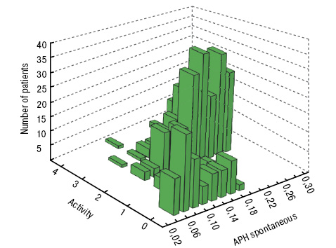
Figure. Dynamics of APH spontaneous depending on the activity of the disease
There was a progressive increasing in the indices, in particular the quantitative characteristics of the oxidative degradation of plasma proteins on the level of PH. Also registered regressing of SOD in eRA with increasing inflammation, but no significant differences in static specified index was achieved when comparing those with 1 and 3, the degree of activity of eRA (–52.41%). Also, due to the large spread, the statistical difference between the KPH, depending on the intensity of the disease was also not recorded, although it should be noted that if eRA patients with minimal activity — this indicator was only 107.31% less, the patients with severe activity-differences were already 216.83%, values of antioxidant activity of this group patients (lower quartile was 15 s.u.) were significantly lower than normal values.
Evolution of the parameters oxidative modification of proteins, according to the radiological stage of eRA, was of mixed: without undergoing significant changes in the two groups at 1 and 2 stages. Differences for the KPH without induction were more pronounced and were –20.37% and -19.63% (p<0.05). Dynamics of serum antioxidant reserve of the subject also proved reliable predictor.
Noted, that patients with eRA augments progressively the severity of violations of redox homeostasis, as eloquently illustrated how the data changes in the structure of proteins initially and after induction of metal, especially the APH and regressing to an intensification of the generation of free radicals in oxidative stress. Further, in order to evaluate the effect of free radical oxidation activation of biomolecules on the dynamics of immune-inflammatory processes and the severity of the bone erosions in eRA, we’ve conducted analysis of variance (ANOVA) (table 2).
| Factor | Dispersion | SS | Degrees of latitude | MS | F | p | η2 | η |
|---|---|---|---|---|---|---|---|---|
| Anti-CCP | intergroup | 725,467 | 1 | 725,467 | 30,193 | <0,001 | 0,335 | 0,579 |
| residual | 1441,638 | 60 | 24,027 | – | – | |||
| general | 2167,105 | 61 | – | – | – | |||
| TNF-α | intergroup | 0,011 | 1 | 0,011 | 15,317 | <0,001 | 0,282 | 0,531 |
| residual | 0,028 | 38 | 0,001 | – | – | |||
| general | 0,039 | 39 | – | – | – | |||
| Larsen Index | intergroup | 4524,745 | 1 | 4524,745 | 10,072 | 0,003 | 0,201 | 0,449 |
| residual | 17 969,146 | 40 | 449,229 | – | – | |||
| general | 22 493,891 | 41 | – | – | – |
SS — sum of squares; MS — the average sum of squares; dl — number of degrees of latitude; F — F-value ratio Fisher; р — reached the significance level of differences, η2 — specific value impact factor underlying the group of a set of factors that affect the effective sign; η –empirical correlation ratio.
The very high value of the F-ratio 30.193 index and the close functional relationship between the APH and anti-CCP, as evidenced by relatively high figure calculated empirical correlation ratio (0.58). Also found a statistically significant effect oxidative modification to a resultant variable as TNF-α. In the corresponding dispersion complex, the reported APH spontaneous reliably account for about 53% of the variation in the values of TNF ηe=0.28 and F=15.32, indicating a relationship between the degree of intensification of carbonyl stress in eRA and the severity of immunopathology viagra high processes. The results of the calculation of the dispersion relations and the proportion of the total variance, which is influenced by APH on the Larson’s index: 10.07 and 20% at p=0.003. Moreover, consideration of the functional dependence by calculation of the empirical correlation ratio (0.45) allowed to establish a close relationship between the studied features.
Oxidative stress acts as one of the leading factors of joint tissue damage in eRA, and the mechanisms are in direct effect, and indirectly, through the activation the inflammatory cascade. The results show, that the patients with eRA have the activation of peroxidation processes, which causes the development of metabolic disorders, changes in the cell membrane and the formation of erosions [11, 13].
Thus, the data obtained allow to conclude, that there is a marked imbalance between eRA activity generating free radicals and their elimination under the influence of endogenous antioxidant systems, characterized by significant prevailing activity of free radicals and oxidative stress status.
ОКСИДАТИВНАЯ МОДИФИКАЦИЯ БЕЛКОВ И КОСТНЫЕ ЭРОЗИИ У БОЛЬНЫХ РАННИМ РЕВМАТОИДНыМ АРТРИТОМ: ЧТО ОБЩЕГО?
Резюме. Цель исследования — определение интенсивности карбонильного стресса при раннем ревматоидном артрите (рРА) с оценкой его патогенетической взаимосвязи с воспалительно-эрозивным процессом. Альдегидные и кетонные формы дигидрофенилгидразонов были оценены в качестве маркеров активации свободных радикалов. Антитела к цитруллинированному пептиду, фактору некроза опухоли-α определяли иммуноферментным анализом. Лица с клинически значимыми сопутствующими заболеваниями или состояниями были тщательно исключены. Обработка и анализ данных проводили с помощью статистических пакетов программ «SPSS 16 для Windows» и «Statistica для Windows 7.0» (StatSoftInc.). Уровень статистической значимости принят р<0,05. Результаты исследования показали, что пациенты с РА характеризуются увеличением свободной генерации радикалов, согласно уровню альдегидных и кетонных формы дигидрофенилгидразонов, ранних маркеров окислительных процессов. Полученные данные позволяют сделать вывод, что у больных рРА при активации воспалительного процесса происходит активация процессов пероксидации в рамках карбонильного стресса, что вызывает развитие дистрофических и деструктивных изменений в хрящевой ткани с нарушением обмена протеогликанов и формирование эрозий.
Ключевые слова: ранний ревматоидный артрит, фенилгидразон, матриксные металлопротеиназы, супероксиддисмутаза.
ОКСИДАТИВНА МОДИФІКАЦІЯ БІЛКІВ ТА КІСТКОВІ ЕРОЗІЇ У ХВОРИХ НА РАННІЙ РЕВМАТОїДНИЙ АРТРИТ: ЩО СПІЛЬНОГО?
Резюме. Мета дослідження — визначення інтенсивності карбонільного стресу при ранньому ревматоїдному артриті (рРА) з оцінкою його патогенетичного взаємозв’язку із запально-ерозивним процесом. Альдегідні й кетонні форми дигідрофенілгідразонів були оцінені в якості маркерів активації вільних радикалів. Антитіла до цитрулінованого пептиду, фактору некрозу пухлини-α визначали імуноферментним аналізом. Особи з клінічно значущими супутніми захворюваннями чи станами були ретельно виключені. Обробку та аналіз даних проводили за допомогою статистичних пакетів програм «SPSS 16.0 для Windows» і «Statistica для Windows 7.0» (StatSoftInc.). Рівень статистичної значущості прийнятий р<0,05. Результати дослідження показали, що пацієнти з РА характеризуються збільшенням вільної генерації радикалів, згідно рівню альдегідних і кетоних форм дигідрофенілгідразонів, ранніх маркерів окисних процесів. Отримані дані дозволяють зробити висновок, що у хворих на рРА при активації запального процесу відбувається активація процесів пероксидації в рамках карбонільного стресу, що викликає розвиток дистрофічних і деструктивних змін у хрящовій тканині з порушенням обміну протеогліканів і формування ерозій.
Ключові слова: ранній ревматоїдний артрит, фенілгідразон, матриксні металопротеїнази, супероксиддисмутаза.
References
1. Мазуров В.И. (ред.) (2005) Клиническая ревматология: руководство для врачей. 2-е изд., перераб. и доп. ФОЛИАНТ, Санкт-Питербург, 520 с.
2. Боброва Л.Н. (1995) Клинико-диагностическое значение определения антител к супероксиддисмутазе. глутатионредуктазе у больных ревматоидным артритом. Дис. … канд. мед. наук, Волгоград, 130 c.
3. Hagfors L., Leanderson P., Skoldstam L. et al. (2003) Antioxidant intake, plasma antioxidants and oxidative stress in a randomized, controlled, parallel, Mediterranean dietary intervention study on patients with rheumatoid arthritis. Nutr. J., 2: 5 (doi: 10.1186/1475–2891–2–5).
4. Taysi S., Polat F., Gul M. et al. (2002) Lipid peroxidation, some extracellular antioxidants, and antioxidant enzymes in serum of patients with rheumatoid arthritis. Rheumatol. Int., 21: 200–204.
5. Зборовская И.А., Баникова М.В. (1994) Патогенетическое и клинико-диагностическое значение показателей антиоксидантной системы и содержания продуктов перекисного окисления липидов у больных ревматическими болезнями. Клин. ревматол., 4, с. 1316.
6. Микунис Р.И. (1989) Состояние антиоксидантов и антиоксидаитной системы у больных ревматоидным артритом. Терапевт. арх., 61(6): 121–123.
7. Носков С.М., Козлов Г.С., Широкова Л.Ю. (1998) Свободнорадикальные реакции при ревматоидномартрите (Обзор). Ревматология, 4: 72–76.
8. Babior B.M. (2000) Phagocytes and oxidative stress. Am. J. Med., 109: 33–44.
9. Cemerski S., van Meerwijk J.P., Romagnoli P. (2003) Oxidative-stress-induced T lymphocyte hyporesponsiveness is caused by structural modification rather than proteasomal degradation of crucial TCR signaling molecules. Eur. J. Immunol., 33: 2178–2185.
10. Мульдияров П.Я., Талыбов Ф.Ю., Николаев В.И. (1993) Состояние свободнорадикалыюго окисления у больных ревматоидным артритом с анемией. Тер. арх., 5, с. 1922.
11. Rees M.D, Hawkins C.L., Davies M.J. (2003) Hypochlorite-mediated fragmentation of hyaluronan, chondroitin sulfates, and related N-acetyl glycosamines: evidence for chloramide intermediates, free radical transfer reactions, and site-specific fragmentation. J. Am. Chem. Soc., 125: 13719–13733.
12. Heliovaara M., Knekt P., Aho K. et al. (1994) Serum antioxidants and risk of rheumatoid arthritis. Ann. Rheum. Dis., 53: 51–53.
13. Henrotin Y.E., Bruckner P., Pujol J.P. (2003) The role of reactive oxygen species in homeostasis and degradation of cartilage. Osteoarthritis. Cartilage, 11: 747–755.
14. Paredes S., Girona J., Hurt-Camejo E. et al. (2002) Antioxidant vitamins and lipid peroxidation in how to drink cialis patients with rheumatoid arthritis: association with inflammatory markers. J. Rheumatol., 29: 2271–2277.
15. Rees M.D, Hawkins C.L., Davies M.J. (2004) Hypochlorite and superoxide radicals can act synergistically to induce fragmentation of hyaluronan and chondroitin sulfates. Biochem. J., 381: 175–184.
16. Dalle-Donne I., Rossi R., Giustarini D. et al. (2003) Protein carbonyl groups as biomarkers of oxidative stress. Clin. Chim. Acta., 329: 23–38.
17. Taysi S., Polat F., Gul M. et al. (2002) Lipid peroxidation, some extracellular antioxidants, and antioxidant enzymes in serum of patients with rheumatoid arthritis. Rheumatol. Int., 21: 200–204.
Correspondence:
Kovalenko Volodymyr Petrovych
03680, Kyiv, Narodnogo opolchenia, 5
National Scientific Center
«M.D. Strazhesko Institute
of Cardiology, NMAS of Ukraine»
Leave a comment